RESPIRATORY SYSTEM
September 21, 2004David S. Phelps, Ph.D.,
Department of Pediatrics, C7814
dsp4@psu.edu
READING – Langman’s Medical Embryology, by T.W. Sadler; 9th edition; 2004.
Chapter 10 – pp. 216-221
Chapter 12 – complete
Chapter 15 – pp. 370-375; 382-3
Learning Objectives
1. Explain how defects in tracheal/esophageal development occur.
2. Describe the embryonic origin of the laryngeal cartilages.
3. Discuss the role of dichotomous branching in the process of lung development
4. SAVE FOR CMBP - Explain the relationship between pulmonary surfactant and neonatal respiratory distress syndrome.
5. Explain how different components participate in the partitioning of the body cavities and the development of the diaphragm.
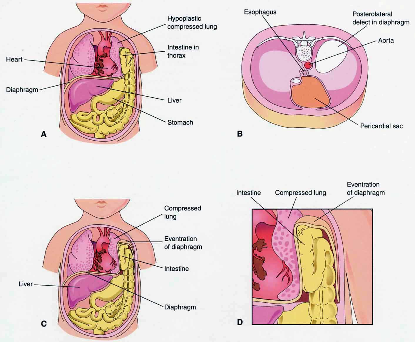
6. Describe how this process is altered in the development of a diaphragmatic hernia.
Most handout figures from Moore and Persaud’s "Before We Are Born" 6th edition.
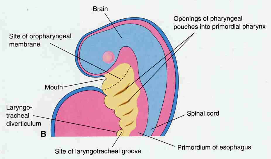
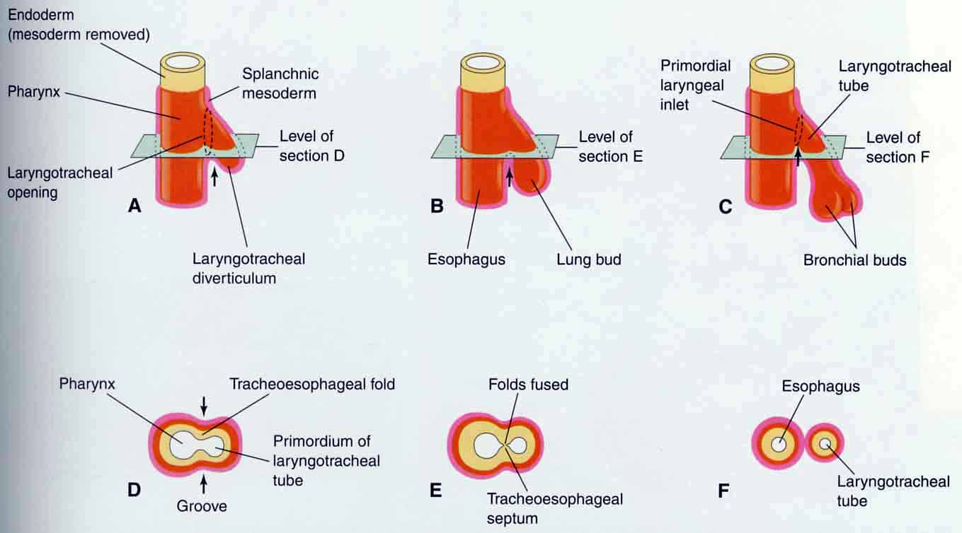
A. Lung bud or laryngotracheal diverticulum
(see also Sadler 12-1)
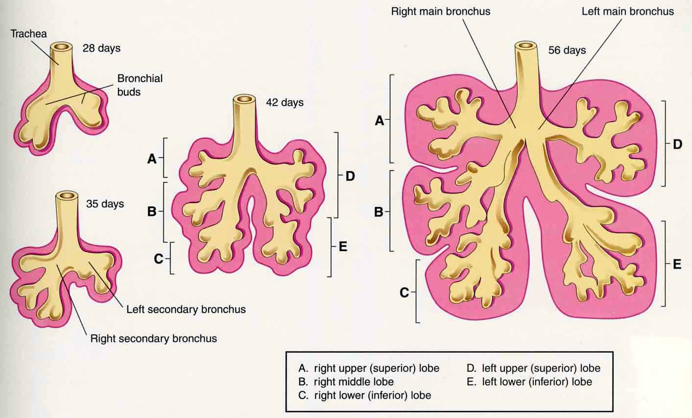
B. Dichotomous branching (see also Sadler 12-2; 12-5)
C. 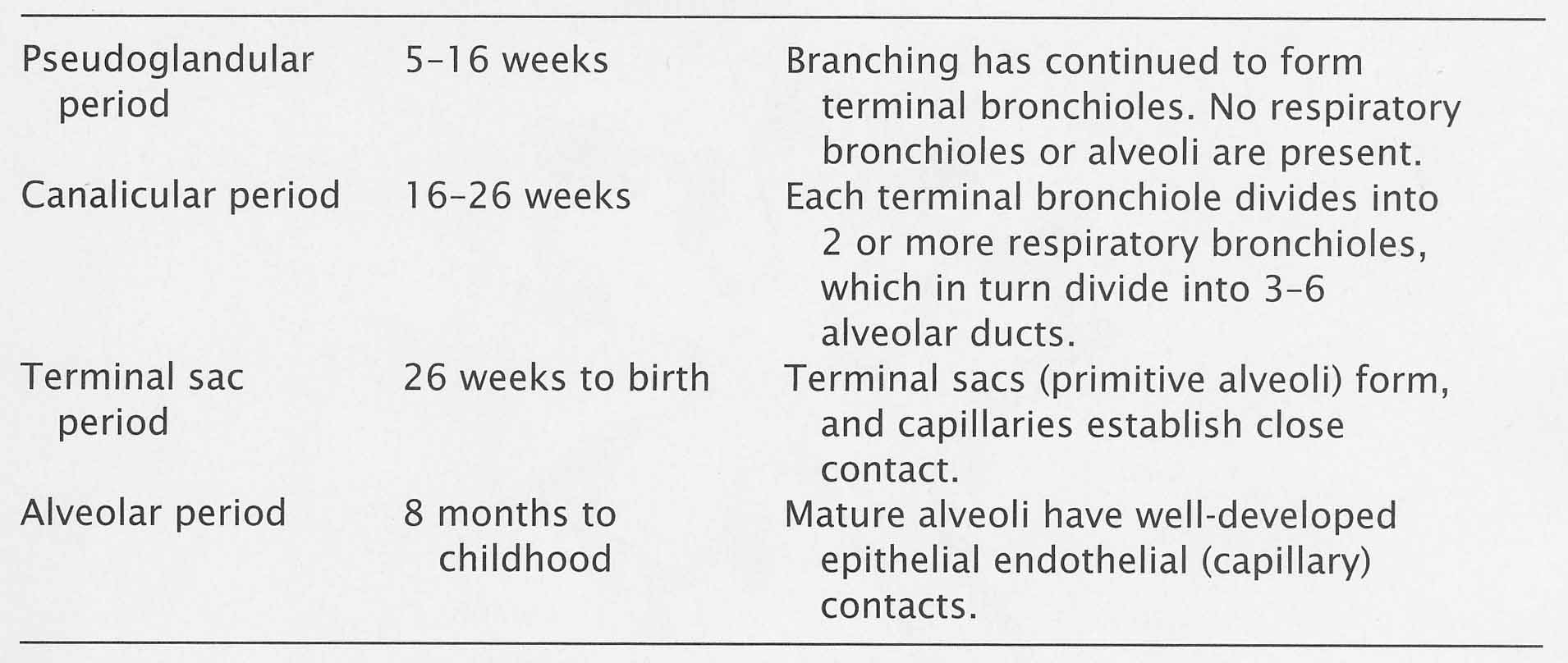
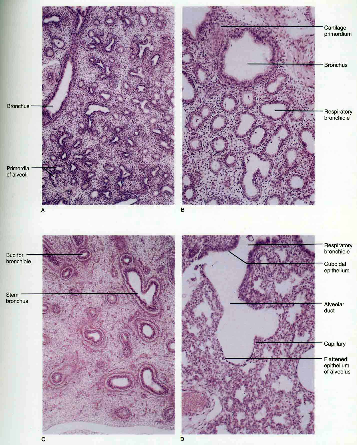 Stages of development (see also Sadler 12-8; 12-9)
Sadler – Table 12.1 Maturation of the Lungs
II. Development of the Larynx
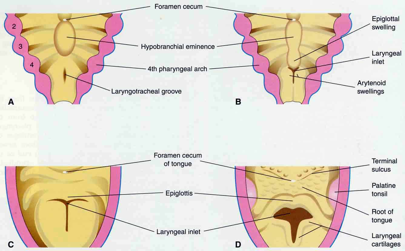
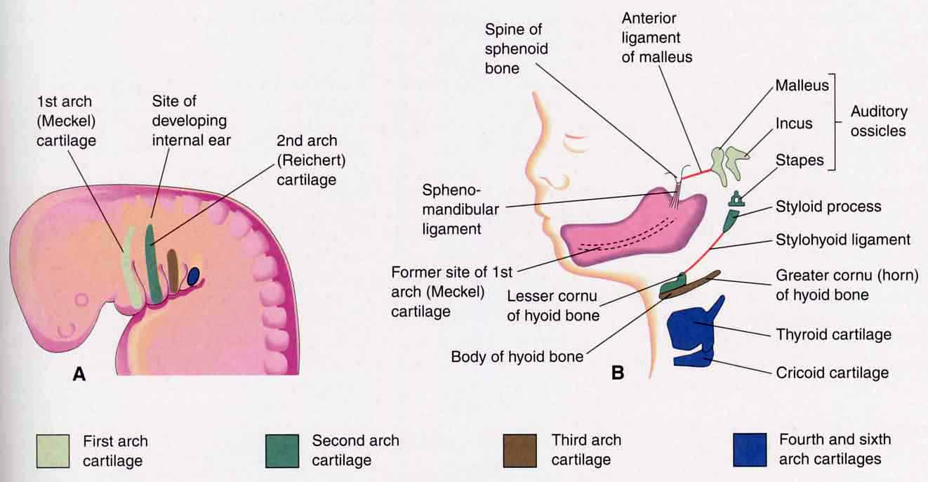
A. Pharyngeal arches and laryngeal cartilages (see also Sadler 12-2; 15-7; 15-8; 15-9).
also Sadler 12-2).
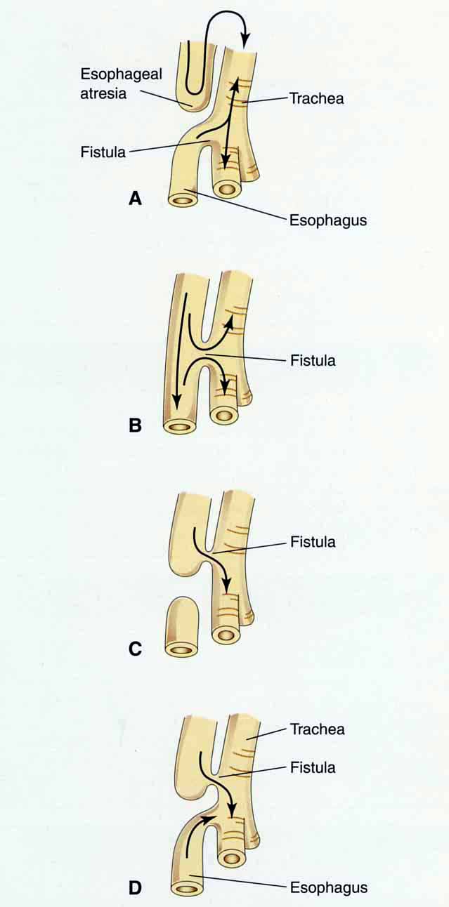
C. Tracheoesophageal malformations (Fig 12.5)
These malformations belong to a group of defects that often occurs together known as the VACTERL association (also VATER or VACTER, depending on author). VACTERL is an acronym that stands for Vertebral, Anal, Cardiac, Trachea, Esophageal, Renal, and Limb anomalies. ). This group may have a common mechanism or timing, but the mechanism has not yet been confirmed. It is possible that there is a connection between this group of anomalies and the use of oral contraceptives early in pregnancy.
III. Diaphragm Development
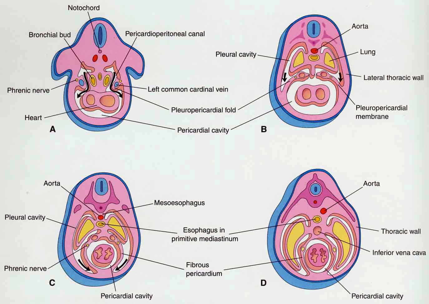
A. Partitioning of pericardial and pleural cavities
(see also Sadler 10-4; 10-5).
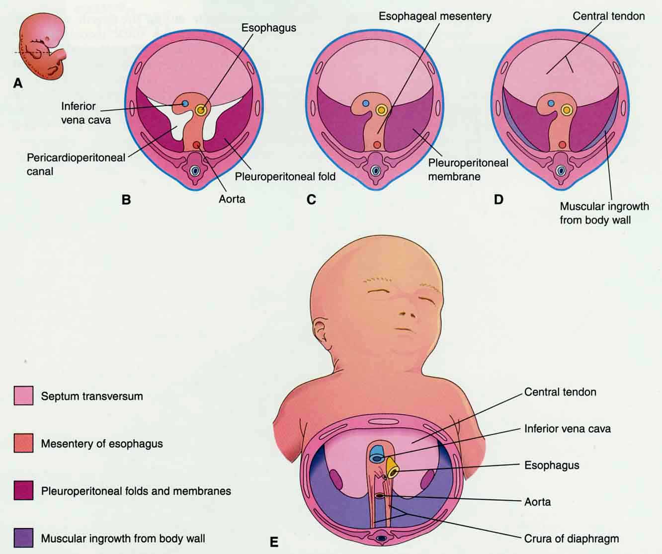
B. Formation of diaphragm (see also Sadler 10-6).
(note orientation)
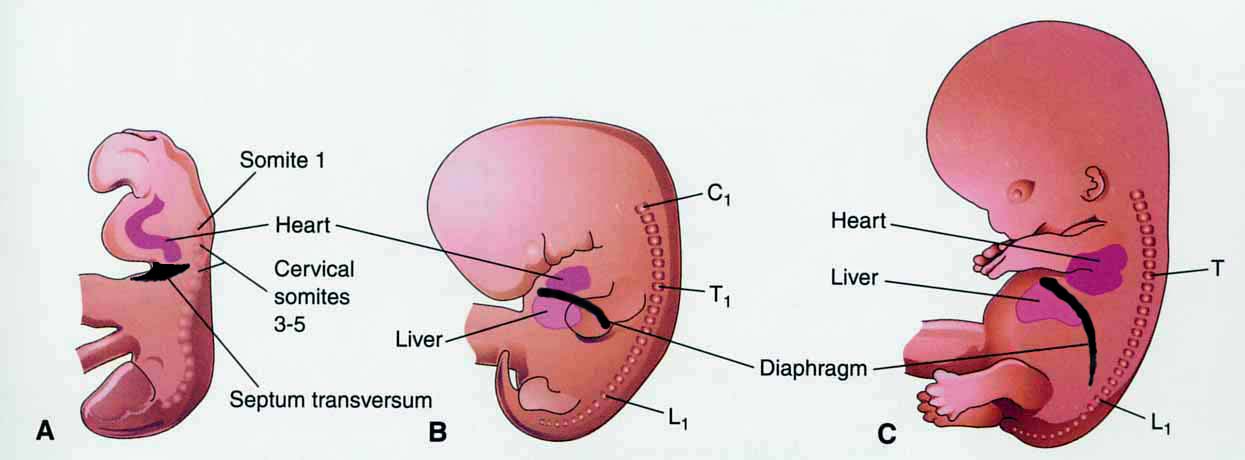
D. Descent of the diaphragm
References
1. Langman’s Medical Embryology, 9th edition. T. W. Sadler. Lippincott, Williams and Wilkins, Philadelphia, 2004.
2. Before We Are Born, 6th edition. K.L. Moore and T.V.N. Persaud. W. B. Saunders Co, Philadelphia, 2003.
3. The Developing Human, 6th edition. K.L. Moore and T.V. N. Persaud. W. B. Saunders, Philadelphia, 1998.
4. Human Embryology and Developmental Biology. B. M. Carlson. Mosby, Philadelphia, 1994.
5. Human Embryology, 3rd edition. W. J. Larsen. Churchill Livingstone, Philadelphia, 2001.